Magnetic Particle Imaging

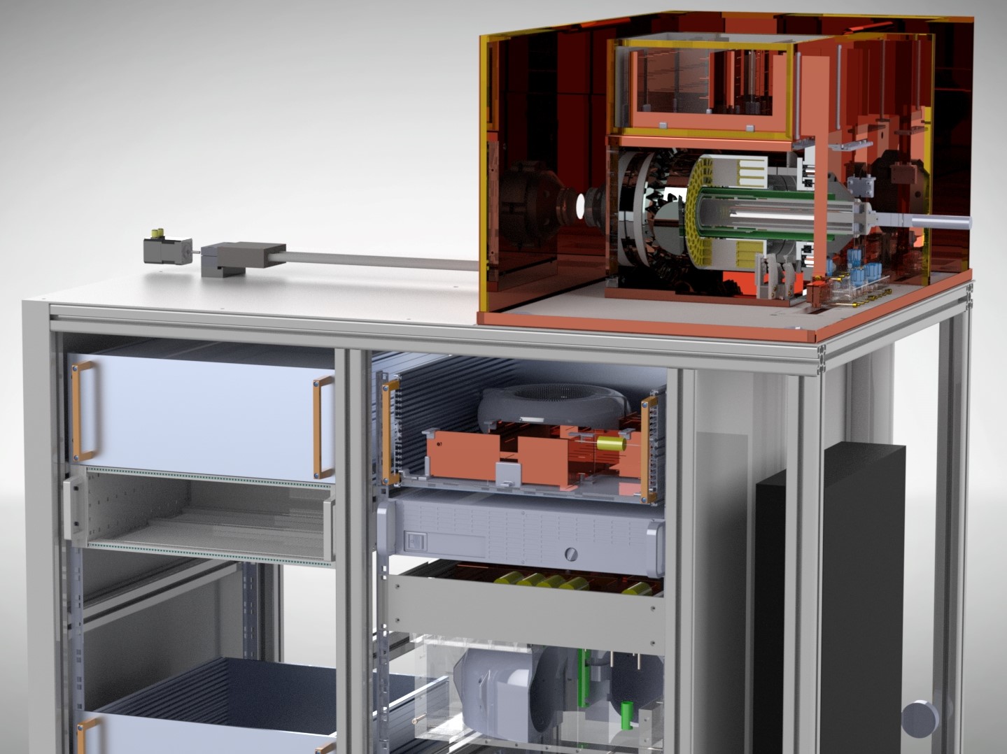
Magnetic Particle Imaging (MPI) is a tomographic imaging modality that allows the quantitative measurement and visualization of distribution and concentration of magnetic nanoparticles with high spatial and temporal resolution and high sensitivity. The method is based on the non-linear magnetization behavior of nanoparticles within homogeneous gradient fields and dynamic alternating fields and opens up the possibility of a radiation-free supplement to existing imaging techniques.

In addition to basic physical and chemical research in the field of magnetic nanoparticles, the basis of this method, research at IMTE focuses on the instrumentation of this medical imaging methodology as well as the image reconstruction.

Find out more about our services regarding Technical and Preclinical Imaging, Magnetic Systems and Instrumentation and Algorithm Design.

Instrumentation

MPI instrumentation comprises the analysis and development of all components necessary for clinical and preclinical imaging along the transmission and reception chain of this imaging modality as well as the development of devices for testing and characterizing nanoparticles, including the identification and consideration of potentials for clinical applications and therapies. Instrumentation is a crucial part of the research to realize the full potential of MPI. This includes the development of dedicated filter circuits and special field topologies to achieve spatial coding and particle excitation. The recording of the weak particle signals with high quality is achieved by the development of amplifiers specially tuned to the receiving characteristics of the respective system.

Research at Fraunhofer IMTE is particularly focused on the combination of electromagnets and permanent magnets for the generation of the required fields. Using a smart arrangement of permanent magnets in a so-called Halbach configuration, strong magnetic fields can be realized without the need for electrical power for field generation. This leads to compact systems which have high potential for a number of applications, especially in preclinical research. Our current scanner concept offers an ideal basis for preclinical research with functionalized magnetic nanoparticles and, to the same extent, for the further development of instrumentation and dedicated reconstruction algorithms.

Reconstruction

The image reconstruction in MPI can be performed in several ways depending on the utilized hardware, tracer material and imaging sequence. At the IMTE, application-specific algorithms are developed for producing optimal image quality for different medical fields and tasks. Research aspects of special interest are for example the identification of new imaging sequences, real-time image reconstruction, enlargement of the imaging volume and the usage of hybrid system matrices to decrease calibration time. Individual adjustment of the algorithms and the reconstruction parameters is necessary to allow optimal usage of MPI's advantages in diagnosis, treatment and therapy.